image segmentation
 With our model for the shape matching
problem, we were capable of segmenting 256 x 256 MRI brain images
using a single level set function (see colored picture below) which
combined with our representation of a continuous Heaviside functional
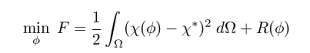
gives sharp segmented areas. This is our first attempt at variational image processing.
With our model for the shape matching
problem, we were capable of segmenting 256 x 256 MRI brain images
using a single level set function (see colored picture below) which
combined with our representation of a continuous Heaviside functional
gives sharp segmented areas. This is our first attempt at variational image processing.
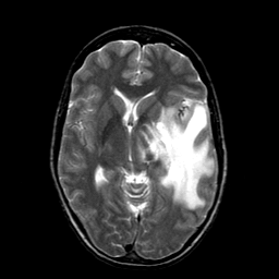
This is one of the original brain images we used to test our shape matching model for image segmentation. It is available at the Whole Brain Atlas, Copyright © 1995-1999 Keith A. Johnson and J. Alex Becker. It shows a case of Metastatic bronchogenic carcinoma, a brain tumor (the large white patch) that affected a 42 year old woman with a long history of tobacco use. This is case 28 in the atlas.
It took only 9 iterations for our nonlinear solver to compute the final
discretized level set function. We have obtained similar number of
iterations for other brain images.
We want to try noisy images using Total Variation and work on
preserving the good efficiency of our optimization algorithms.
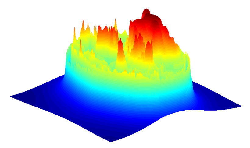
This picture shows the final level set function obtained using our optimization algorithm. It is a C1 differentiable smooth function defined over a triangular mesh according to our finite element approximation. Note that it is far from being a signed distance function.
Click on image to see a larger version.
Below is the sequence of contours extracted from the same evolving level set function, two per frame, both drawn on top of the target image. The starting level set function is a circle centered at the image. Each frame represents one iteration of the nonlinear solver starting from initial configuration (frame 0). The black contour corresponds to the zero level set and the red contour surrounds areas for which the pixel value is greater or equal to 250.
An animation of these pictures is also available. Check also an animation of the images generated at each iteration with the contours.
0 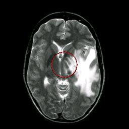 |
1 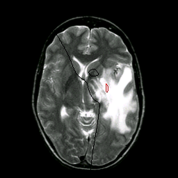 |
2 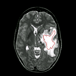 |
3 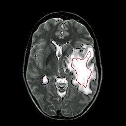 |
4 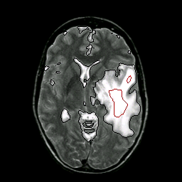 |
5 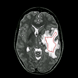 |
6  |
7 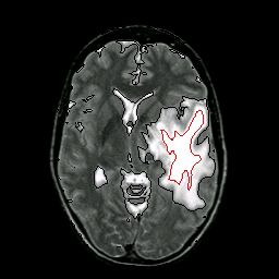 |
8  |
9 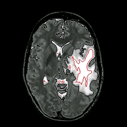 |
Copyright Alexandre Cunha
updated on jul/2004